
Facilitating rapid discovery and development of potentially predictive signatures with the most advanced view of immuno-oncology biology.
The PanCancer IO 360 Gene Expression Panel...
PanCancer Pathways
nCounter PanCancer Pathways Panel
Drawing on 30+ years of cancer research, QPCR, Next Generation Sequencing, Gene Expression, Western blotting the cancer community has accomplished a lot to narrow tens of thousands of genes to the ones that matter for the study of cancer.
Today, we are taking the next step in that journey with the introduction of our PanCancer Pathways Panel, a novel set of 700 essential genes representing all major cancer pathways including key driver genes.
Using a biology-guided, data-driven approach and applying basic co-expression principles we scored and ranked each gene based on its biological relevance to cancer and its role as an essential representation in one or more of the 13 canonical pathways.
We can now take this very complex disease and make it a bit easier to comprehend to
Discover connections. Discover interactions. Discover function.
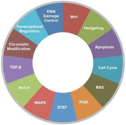
Pathways of the PanCancer Pathways Panel
770 Essential Genes Representing 13 Canonical Pathways
- 606 Pathway Genes:
Notch, Wnt, Hedgehog, TGFB, MAPK, STAT, P13K, RAS, Chromatin Modification, Transcriptional Regulation, DNA Damage Control, Cell Cycle, Apoptosis - 124 cancer driver genes
- 40 reference genes
- Customize with Panel-Plus option flexibility to include 30 genes of your choice
Below are brief descriptions of the Pathways included in the PanCancer Pathways Panel. Please click on the name of the pathway to view more information, including detailed information on pathway genes and KEGG pathway gene maps.
|
Pathway |
Description |
|
Notch |
The Notch signaling pathway is an evolutionarily conserved, intercellular signaling mechanism essential for proper embryonic development. The Notch proteins are single-pass receptors that are activated by the Delta (or Delta-like) and Jagged/Serrate families of membrane-bound ligands. They are transported to the plasma membrane as cleaved, but otherwise intact polypeptides. Interaction with ligand leads to two additional proteolytic cleavages that liberate the Notch intracellular domain (NICD) from the plasma membrane. The NICD translocates to the nucleus, where it forms a complex with the DNA binding protein CSL, displacing a histone deacetylase (HDAc)-co-repressor (CoR) complex from CSL. Components of an activation complex, such as MAML1 and histone acetyltransferases (HATs), are recruited to the NICD-CSL complex, leading to the transcriptional activation of Notch target genes. |
|
APC (Wnt) |
Wnt proteins are secreted morphogens that are required for basic developmental processes, such as cell-fate specification, progenitor-cell proliferation and the control of asymmetric cell division, in many different species and organs. There are at least three different Wnt pathways: the canonical pathway, the planar cell polarity (PCP) pathway and the Wnt/Ca2+ pathway. In the canonical Wnt pathway, the major effect of Wnt ligand binding to its receptor is the stabilization of cytoplasmic beta-catenin through inhibition of the bea-catenin degradation complex. Beta-catenin is then free to enter the nucleus and activate Wnt-regulated genes through its interaction with TCF (T-cell factor) family transcription factors and concomitant recruitment of coactivators. Planar cell polarity (PCP) signaling leads to the activation of the small GTPases RHOA (RAS homologue gene-family member A) and RAC1, which activate the stress kinase JNK (Jun N-terminal kinase) and ROCK (RHO-associated coiled-coil-containing protein kinase 1) and leads to remodeling of the cytoskeleton and changes in cell adhesion and motility. WNT-Ca2+ signaling is mediated through G proteins and phospholipases and leads to transient increases in cytoplasmic free calcium that subsequently activate the kinase PKC (protein kinase C) and CAMKII (calcium calmodulin mediated kinase II) and the phosphatase calcineurin. |
|
Hedgehog |
The Hedgehog (Hh) family of secreted signaling proteins plays a crucial role in development, regulating morphogenesis of a variety of tissues and organs. Hh signaling is also involved in control of stem cell proliferation in adult tissues and aberrant activation of the Hh pathway has been linked to multiple types of human cancer. Members of the Hh family bind to patched (ptc), thus releasing smoothened (smo) to transduce a signal. Transcriptional activation occurs through the GLI family of proteins resulting in activation of target genes. |
|
Chromatin Modification |
Members of this family of genes are involved or regulate processes associated with the alteration of DNA, protein, or sometimes RNA, in chromatin, which may result in changing the chromatin structure. |
|
Transcriptional Regulation |
A collection of pathways annotated by KEGG known to be transcriptionally misregulated in a variety of cancers. |
|
DNA Damage Control |
DNA repair is a multi-enzyme, multi-pathway system required to ensure the integrity of the cellular genome. DNA damage can arise spontaneously in the cellular milieu through chemical alteration of base nucleotides or as a consequence of errors during DNA replication. The basic mechanisms underlying distinct DNA repair pathways include nucleotide excision repair (NER), base excision repair (BER), DNA strand break repair (DSBR), direct reversal of DNA damage, and the replication past DNA lesions by specialized DNA bypass polymerases (bypass replication). Defects in most of these repair pathways have been associated with one or more specific human diseases. Additionally, the repair of damaged DNA is intimately associated with a number of other distinct cellular processes such as DNA replication, DNA recombination, cell cycle checkpoint arrest, and other basic cellular mechanisms. |
|
TGF-β |
The transforming growth factor-beta (TGF-beta) family members, which include TGF-betas, activins and bone morphogenetic proteins (BMPs), are structurally related secreted cytokines. A wide spectrum of cellular functions such as proliferation, apoptosis, differentiation and migration are regulated by TGF-beta family members. TGF-beta family member binds to the Type II receptor and recruits Type I, whereby Type II receptor phosphorylates and activates Type I. The Type I receptor, in turn, phosphorylates receptor-activated Smads ( R-Smads: Smad1, Smad2, Smad3, Smad5, and Smad8). Once phosphorylated, R-Smads associate with the co-mediator Smad, Smad4, and the heteromeric complex then translocates into the nucleus. In the nucleus, Smad complexes activate specific genes through cooperative interactions with other DNA-binding and coactivator (or co-repressor) proteins. |
|
MAPK |
The mitogen-activated protein kinase (MAPK) cascade is a highly conserved module that is involved in various cellular functions, including cell proliferation, differentiation and migration. Mammals express at least four distinctly regulated groups of MAPKs, extracellular signal-related kinases (ERK)-1/2, Jun amino-terminal kinases (JNK1/2/3), p38 proteins (p38alpha/beta/gamma/delta) and ERK5, that are activated by specific MAPKKs: MEK1/2 for ERK1/2, MKK3/6 for the p38, MKK4/7 (JNKK1/2) for the JNKs, and MEK5 for ERK5. Each MAPKK, however, can be activated by more than one MAPKKK, increasing the complexity and diversity of MAPK signaling. |
|
STAT |
The Janus kinase/signal transducers and activators of transcription (JAK/STAT) pathway is one of a handful of pleiotropic cascades used to transduce a multitude of signals for development and homeostasis in animals. In mammals, the JAK/STAT pathway is the principal signaling mechanism for a wide array of cytokines and growth factors. Following the binding of cytokines to their cognate receptor, STATs are activated by members of the JAK family of tyrosine kinases. Once activated, they dimerize and translocate to the nucleus and modulate the expression of target genes. In addition to the activation of STATs, JAKs mediate the recruitment of other molecules such as the MAP kinases, PI3 kinase etc. These molecules process downstream signals via the Ras-Raf-MAP kinase and PI3 kinase pathways which results in the activation of additional transcription factors. |
|
PI3K |
The phosphatidylinositol 3' -kinase(PI3K)-Akt signaling pathway is activated by many types of cellular stimuli or toxic insults and regulates fundamental cellular functions such as transcription, translation, proliferation, growth, and survival. The binding of growth factors to their receptor tyrosine kinase (RTK) or G protein-coupled receptors (GPCR) stimulates class Ia and Ib PI3K isoforms, respectively. PI3K catalyzes the production of phosphatidylinositol-3,4,5-triphosphate (PIP3) at the cell membrane. PIP3 in turn serves as a second messenger that helps to activate Akt. Once active, Akt can control key cellular processes by phosphorylating substrates involved in apoptosis, protein synthesis, metabolism, and cell cycle. |
|
RAS |
The Ras proteins are GTPases that function as molecular switches for signaling pathways regulating cell proliferation, survival, growth, migration, differentiation or cytoskeletal dynamism. Ras proteins transduce signals from extracellular growth factors by cycling between inactive GDP-bound and active GTP-bound states. The exchange of GTP for GDP on RAS is regulated by guanine nucleotide exchange factors (GEFs) and GTPase-activating proteins (GAPs). Activated RAS (RAS-GTP) regulates multiple cellular functions through effectors including Raf, phosphatidylinositol 3-kinase (PI3K) and Ral guanine nucleotide-dissociation stimulator (RALGDS). |
|
Cell Cycle |
Mitotic cell cycle progression is accomplished through a reproducible sequence of events, DNA replication (S phase) and mitosis (M phase) separated temporally by gaps known as G1 and G2 phases. Cyclin-dependent kinases (CDKs) are key regulatory enzymes, each consisting of a catalytic CDK subunit and an activating cyclin subunit. CDKs regulate the cell's progression through the phases of the cell cycle by modulating the activity of key substrates. Downstream targets of CDKs include transcription factor E2F and its regulator Rb. Precise activation and inactivation of CDKs at specific points in the cell cycle are required for orderly cell division. Cyclin-CDK inhibitors (CKIs), such as p16Ink4a, p15Ink4b, p27Kip1, and p21Cip1, are involved in the negative regulation of CDK activities, thus providing a pathway through which the cell cycle is negatively regulated. Eukaryotic cells respond to DNA damage by activating signaling pathways that promote cell cycle arrest and DNA repair. In response to DNA damage, the checkpoint kinase ATM phosphorylates and activates Chk2, which in turn directly phosphorylates and activates p53 tumor suppressor protein. p53 and its transcriptional targets play an important role in both G1 and G2 checkpoints. ATR-Chk1-mediated protein degradation of Cdc25A protein phosphatase is also a mechanism conferring intra-S-phase checkpoint activation. |
|
Apoptosis |
Apoptosis is a genetically controlled mechanisms of cell death involved in the regulation of tissue homeostasis. The 2 major pathways of apoptosis are the extrinsic (Fas and other TNFR superfamily members and ligands) and the intrinsic (mitochondria-associated) pathways, both of which are found in the cytoplasm. The extrinsic pathway is triggered by death receptor engagement, which initiates a signaling cascade mediated by caspase-8 activation. Caspase-8 both feeds directly into caspase-3 activation and stimulates the release of cytochrome c by the mitochondria. Caspase-3 activation leads to the degradation of cellular proteins necessary to maintain cell survival and integrity. The intrinsic pathway occurs when various apoptotic stimuli trigger the release of cytochrome c from the mitochondria (independently of caspase-8 activation). Cytochrome c interacts with Apaf-1 and caspase-9 to promote the activation of caspase-3. Recent studies point to the ER as a third subcellular compartment implicated in apoptotic execution. Alterations in Ca2+ homeostasis and accumulation of misfolded proteins in the ER cause ER stress. Prolonged ER stress can result in the activation of BAD and/or caspase-12, and execute apoptosis. |
-

-
The Hallmarks of Cancer are one of the most widely recognized organizing principles for the holistic study of cancer. Its profoundly simple framework was first introduced in 2000 b...
-
Perform multiplex gene expression analysis with 770 genes from 24 different immune cell types, common checkpoint inhibitors, CT antigens, and genes covering both the adaptive and i...
-
Perform multiplex gene expression analysis with 770 genes from each step in the cancer progression process including: angiogenesis, extracellular matrix remodeling (ECM), epithelia...
-
A Comprehensive Set of Fusion Genes The nCounter Leukemia Fusion Gene Expression Assay Kit allows researchers to profile a comprehensive set of fusion genes which result from balan...
-
nCounter Vantage™ Gene Fusion Panels Multiplexed Gene Fusion Detection Simplified. Save time and sample material with multiplexed gene fusion detection in a single tube. Detect ra...